NIMBUS
Non-Invasive neuroModulation of deep Brain structures for Upper limb recovery after Stroke
A multicenter study investigating transcranial temporal interference stimulation (tTIS) for stroke recovery
Stroke is a leading cause of long-term disability, and persistent upper-limb impairment remains common despite advances in acute care and rehabilitation. Recovery depends on neuroplastic changes across distributed brain networks, including the striatum, a deep brain structure involved in motor learning and functional recovery.
Recent advances in transcranial temporal interference stimulation (tTIS) now make it possible to modulate deep brain structures non-invasively, opening new possibilities for targeted neurorehabilitation. Previous studies have demonstrated the feasibility and safety of striatal tTIS and have shown improvements in motor learning and changes in activity within striatal networks.
NIMBUS will investigate whether personalised striatal tTIS combined with intensive upper-limb rehabilitation can improve recovery after stroke. The project will conduct a multicentre, randomized, double-blind, placebo-controlled clinical trial involving 84 chronic stroke patients across three rehabilitation centres in Switzerland.
Participants will receive either active or placebo stimulation in combination with rehabilitation therapy. In addition to motor recovery, the study will evaluate effects on daily-life functioning, cognition, mood, and brain network organisation using clinical assessments, magnetic resonance imaging (MRI), electroencephalography (EEG), and combined EEG with transcranial magnetic stimulation (TMS).

NIMBUS will provide the first clinical evaluation of personalised non-invasive stimulation of the striatum as an adjunct to stroke rehabilitation.

A closer look at the science behind the tools:
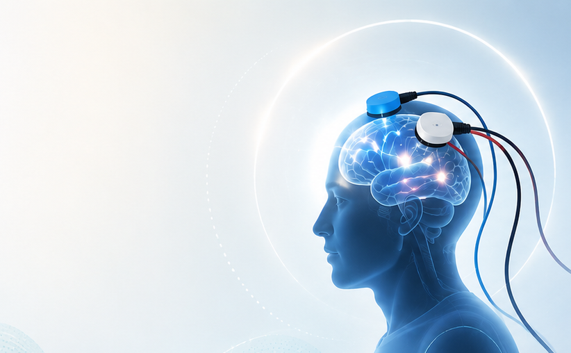
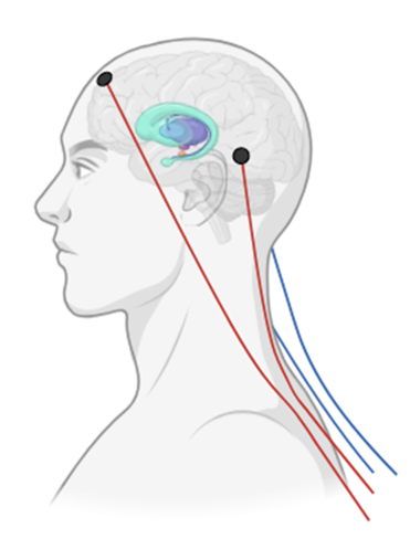
tTIS: A non-invasive method that modulates the activity of deep brain structures safely and painlessly. Two pairs of electrodes placed on the scalp deliver two sources of weak electrical currents through the skull toward the center of the brain; in this particular case, at the striatum.
MRI: A non-invasive imaging method that records brain structures and their functions. It allows to obtain highly detailed images of the brain’s structure and its activity.
EEG: A non-invasive recording method that measures the electrical activity of the brain. It allows to capture real-time brainwave patterns and monitor rapid changes in brain function.
TMS-EEG: A multimodal non-invasive method that combines TMS (which uses magnetic fields to gently stimulate specific areas of the brain) with EEG to study the brain’s reactivity. TMS is used to stimulate the brain, while EEG records exactly how it responds to that stimulation in real time.
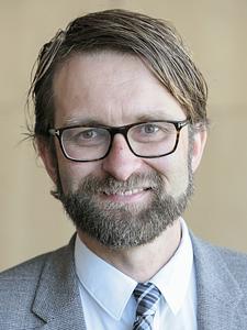


Lucerne Cantonal Hospital (LUKS)

Principal Investigator – Prof. Dr. Thomas Nyffeler

Site coordinator – Gerda Wyssen, Scientist

Leading Therapist – André Hürlimann
Geneva University Hospitals (HUG)

Principal Investigator – Prof. Dr. Adrian Guggisberg